
2D echocardiography, popularly called 2D echo, is a non-invasive test used to analyze the functioning and assess the sections of your heart. This test gives images of the different parts of the heart with the help of sound vibrations.It assists in checking damages, blockages, and blood flow rate.
Doctors recommend regular 2D echo tests to analyze and treat any heart issues at the early stages, keeping you healthy and active as you grow old.
Why should you undergo a 2D echo?
2D Echo is done to detect the following heart conditions:
- Any underlying heart diseases or abnormalities
- Congenital heart diseases and blood clots or tumors
- Malfunctioning of the heart valve
- Abnormality of blood flow within the heart
2D echo gives information related to the functioning of your heart, diagnoses malfunctions, and plans the treatment for the developing disease.
Not only for your doctor but regular 2D echo checkups also help your mind be at peace.
Book your 2D echo test today and see how trading your favorite fast food for a healthy diet works for your heart’s health.
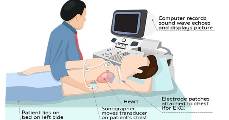
What happens during the 2D Echo Test?
The 2D Echo test takes about an hour. You will be asked to remove any metal jewellery. After that, small adhesive electrodes will be attached to your chest. The electrodes are connected to an ECG machine. A gel will be applied to your chest and then a transducer will be placed and moved around the chest area to get the image of the heart.
What does a 2D echo test show?
The 2D Echo test produces images of different parts of the heart on a computer screen, which is later evaluated by a cardiologist to check for any damage or blockage of the heart tissues and valves. It also evaluates the efficiency of the heart muscles that pump blood to different organs.